Odd silver w/purple inside fruiting body

Evan Sussman

Participating member
Location
Stanwood, WA
This fruiting body was found on the trunk of a decaying but still alive alder tree in Western Washington. Any idea what the fungus is?

P.S. the damage to the fruiting body is from my finger. It's almost chalky inside.
 

Attachments

  • 329549-fungus1.webp
    329549-fungus1.webp
    66.1 KB · Views: 73
I have seen similar to those on my sugar maples in the Adirondacks. It was inside the trunk cavity. I am interested to see what they are. I will look for the pic I took.
 
I shouldn't try to ID stuff from snapshots, but looks to be an aethalium of a myxomycete such as a species of the genus Lycogala. That's a large genus distributed worldwide. Consequently, you might have trouble getting a field-guide picture to line up exactly with what you have.

The characteristics are the smooth, shiny outer wall (peridium) and the chalky dark interior (spore mass). Unlike fungal spores that usually produce hyphae when they germinate, Myxo spores usually produce myxamoebae upon germination, little amoeba-like guys that move around. That's what puts it in with the protists rather than the fungi.

To ID which Myxo you've got, use a microscope. I'd look for capillitia (or psuedocapillitia), fine hairs that help the "timed-release" of the spores. The shape of those hairs are usually good to get it at least to genus.

As a Myxo, it is not a fungus in the modern concept, but a protist. Still, mycologists have traditionaly studied them, so I've poked at them a bit.

Probably though, what you really want to know is "is it damaging the tree?" The answer to that is "no".
 
Hello Kevin,

Thank you for the insightful post. I'm pleased to meet such educated help.

A brief survey of the terminology in your post has been quite educational.

I don't currently have access to a microscope, and the reason for identification is for personal interest. I'm going to ask around though to use one.

How much would I need to be able to magnify the sample to be able to see the capillitia that you speak of?

I have learned that clearly there is much more to identifying a fungus, or protist then brief visual inspection.
 
Welcome to Treebuzz. I learn a lot popping in from time-to-time.

The following is more info than I usually give out, not because I'm secretive but most folks frankly are just not that interested. but here goes:

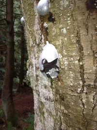
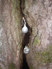
If you have access, say a library copy, of a Myxomycete text, check out Lycogala flavofuscum and see if that fits on a macro basis. That might be all you need.

If you really need to rigorously ID it, a microscope at
100x would likely be enough magnification. if the optics are good enough to provide a sharp image. Resolving power is at least as important as magnification, but it would need to be more than a toy. I'd probably start with *small* amounts of sample on a microscope slide, mounted in water. I'd also probably make a slide with cotton blue in lactophenol and one in Melzer's reagent. I'd be looking for spore ornamentation (pits, spines, warts) and sometimes the dyes help in showing that stuff. If you're not used to doing that stuff or don't know what I'm talking about, then your need is beyond what Treebuzz can provide. Even at 100x, the spores will be quite small, but you should be able to see the diagnostic ornamentation. The pseudocapillitia of Lycogala are like wide, empty tubes as distinct from true capillitia that are usually narrow and forked/branched. Again, the above is routine for a diagnostic lab, but would be overkill for most arborist applications. But like I say, you asked! Happy to help!
 
Thank you,

I must admit that I'm not used to "doing that stuff". However I am intrigued by the study of the Creation.

I found this that I believe to be a Myxomycete text:
http://www.archive.org/stream/myxomycetesdescr00macb/myxomycetesdescr00macb_djvu.txt

Though only the name Lycogala flavofuscum is mentioned. My guess is that the text you are referring to is more in depth.

While searching I found a description of L. epidendrum which looks pretty similar: http://www.messiah.edu/Oakes/fungi_on_wo...0epidendrum.htm


The identification key at :http://www.messiah.edu/Oakes/fungi_on_wood/ while may be basic seems like it could be a useful resource for someone in the Northeast states. What do you think?

From the introduction: Fungi Growing on Wood is a web site devoted to the identification and appreciation of fungi growing in close association with living and dead wood. The fungi described here are those in a parasitic or saprophytic relationship with trees. Mycorrhizal fungi, also in close association with trees, are not strongly represented here even though some of them may be found on well-rotted wood. The organisms described on this web site are often referred to as lignicolous fungi. Most species are illustrated with multiple images to show as many diagnostic details as possible. To my knowledge, no guide similar to this exists.
 
Unfortunately, most field guides don't go too far in depth on the Myxo's. Macbride's monograph (1939) for which you provide a link is good, but mostly cites stuff pre-1925. The descriptions are good, though.

I use "The Myxomycetes" by G.W. Martin, C.J. Alexopoulus, and R.M. Allen (1969) which has good illustrations. *But* is not a field guide or oriented to be useful for the novice. Sorry about that, but some of this stuff takes the equivalent of a few college or graduate school courses to wade through.

There is "Myxomycetes: A Handbook of Slime Molds" by S.L. Stephenson and others (2000 or so) that might be just the ticket, but I haven't used it.

As for online sources, the ones you mention are fine and do satisfy casual interest, but are inadequate for serious work. I'm no Myxo expert, but did hit them pretty well in graduate school.
 

New threads New posts

Back
Top Bottom